Cellular transport malfunctions frequently lead to diseases, including metabolic disorders, hypertension, and organ damage; understanding these processes is crucial for health.
Cellular transport is fundamental to life, governing the movement of substances across cell membranes. This intricate process ensures cells receive essential nutrients while expelling waste products, maintaining internal homeostasis. It’s not merely about entry and exit; it’s a highly regulated system vital for cellular function.
Disruptions in transport mechanisms can trigger a cascade of health issues, ranging from metabolic imbalances to severe organ damage. Understanding these processes is paramount for comprehending disease pathology and developing effective therapeutic strategies. The ability to translate languages, like with Google Translate, mirrors the cell’s need to ‘translate’ external signals into internal responses.
From simple diffusion to complex vesicular transport, cells employ diverse methods to manage their internal environment. This study guide will explore these mechanisms, highlighting their importance and potential vulnerabilities, ultimately revealing how cellular transport underpins overall health and well-being.

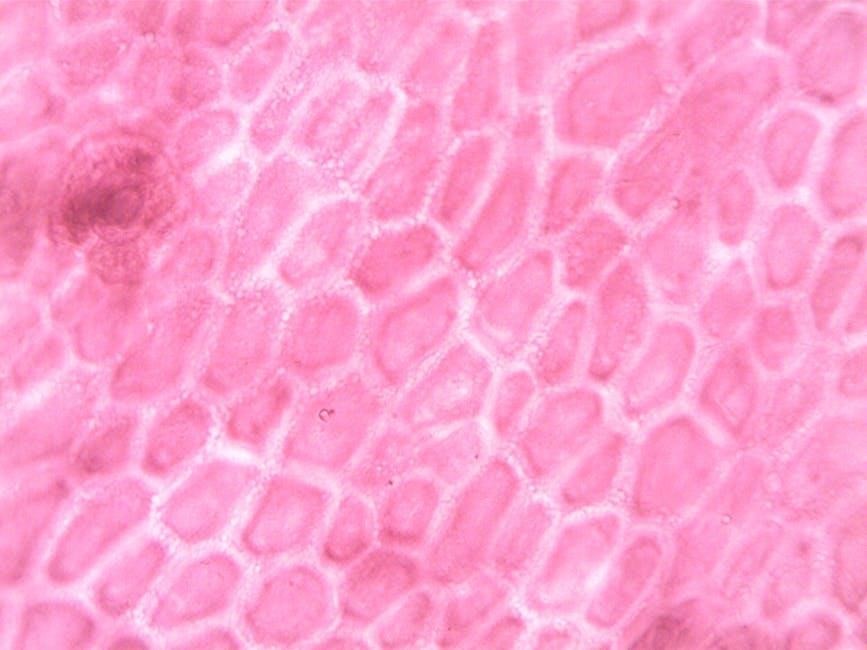
The Plasma Membrane: A Selective Barrier
The plasma membrane acts as a gatekeeper, meticulously controlling the passage of substances into and out of the cell. Its structure, a phospholipid bilayer embedded with proteins, dictates its selective permeability. This barrier isn’t impenetrable; rather, it’s selectively permeable, allowing some molecules to cross easily while restricting others.
This selectivity is crucial for maintaining cellular homeostasis. Like Google Translate facilitating communication between languages, the membrane ‘translates’ external conditions into a suitable internal environment. Transport proteins are key to this process, acting as channels or carriers to facilitate movement.
The membrane’s fluidity and the composition of its lipid and protein components influence its function. Understanding its architecture is essential for grasping how cellular transport operates, and how disruptions can lead to disease, mirroring the impact of inaccurate translations.
Passive Transport Mechanisms
Passive transport relies on the concentration gradient, moving substances across the membrane without expending cellular energy – much like a natural flow. This includes processes like simple diffusion, where molecules move from areas of high to low concentration, and facilitated diffusion, which utilizes transport proteins to assist in this movement.
Osmosis, a special case of diffusion, focuses on water movement across a semi-permeable membrane, driven by water potential differences. Maintaining proper water balance is vital for cell function, similar to ensuring accurate translations in Google Translate for clear communication.
These mechanisms are fundamental for nutrient uptake and waste removal, ensuring cellular survival. Disruptions in passive transport can lead to imbalances, contributing to various health issues, highlighting the importance of a functional ‘gatekeeper’.
Simple Diffusion
Simple diffusion is the most basic means of passive transport, driven solely by the concentration gradient of a substance. Molecules naturally move from an area of high concentration to one of low concentration, requiring no assistance from proteins or energy expenditure – a truly spontaneous process.
This method is effective for small, nonpolar molecules like oxygen and carbon dioxide, allowing them to readily cross the phospholipid bilayer of the cell membrane. Think of it like translating a simple phrase with Google Translate; it’s direct and requires minimal processing.

However, polar and charged molecules face difficulty traversing the hydrophobic core, necessitating alternative transport mechanisms. The rate of diffusion is influenced by temperature, molecule size, and the membrane’s properties.
Facilitated Diffusion
Facilitated diffusion assists the movement of substances across the cell membrane that cannot easily pass through on their own, still adhering to the concentration gradient and remaining a passive process. This relies on the aid of transport proteins – channel or carrier proteins – to expedite the crossing.
Channel proteins create a pore allowing specific molecules or ions to flow through, much like a dedicated translation lane in Google Translate for specific languages. Carrier proteins bind to the substance, changing conformation to shuttle it across, resembling a more complex, nuanced translation.
While requiring proteins, no cellular energy is consumed. This is vital for transporting larger polar molecules like glucose and amino acids, ensuring efficient cellular function.
Osmosis and Water Balance
Osmosis, a special case of diffusion, focuses on water movement across a semi-permeable membrane from an area of high water concentration to low, striving for equilibrium. This is crucial for maintaining cellular volume and function, mirroring Google Translate’s ability to handle diverse linguistic inputs.
Water potential, influenced by solute concentration and pressure, dictates the direction of osmosis. Hypotonic solutions (lower solute) cause water to enter cells, potentially leading to swelling or bursting. Hypertonic solutions (higher solute) draw water out, causing shrinkage. Isotonic solutions maintain balance.
Cells employ mechanisms to regulate water balance, preventing detrimental effects. Understanding osmosis is fundamental to comprehending cellular physiology and its impact on overall health, similar to understanding the core principles of accurate translation.
Active Transport Mechanisms
Active transport requires energy, typically ATP, to move substances against their concentration gradients – from low to high concentration. This contrasts with passive transport, which relies on diffusion and doesn’t need energy input, much like Google Translate utilizing computational power for complex tasks.
Two main types exist: primary and secondary active transport. Primary active transport directly uses ATP, like the sodium-potassium pump, maintaining cellular gradients. Secondary active transport utilizes the electrochemical gradient established by primary transport to move other substances.
These mechanisms are vital for nutrient absorption, waste removal, and maintaining cellular homeostasis. Disruptions in active transport can lead to various diseases, highlighting its importance in overall health and cellular function;
Primary Active Transport
Primary active transport directly utilizes metabolic energy, such as ATP hydrolysis, to fuel the movement of molecules across the cell membrane against their concentration gradient. A classic example is the sodium-potassium pump (Na+/K+ ATPase), crucial for maintaining electrochemical gradients essential for nerve impulse transmission and cellular volume regulation.
This pump actively transports three sodium ions out of the cell for every two potassium ions pumped in, requiring one ATP molecule per cycle. Other examples include the calcium pump and proton pumps, all directly coupled to ATP breakdown.
These processes are fundamental for establishing and maintaining cellular gradients, enabling numerous other cellular functions and demonstrating the cell’s energy expenditure for vital transport.

Secondary Active Transport
Secondary active transport leverages the electrochemical gradient established by primary active transport to move other molecules across the cell membrane. It doesn’t directly use ATP; instead, it relies on the potential energy stored in the gradient of ions like sodium or chloride.
There are two main types: symport, where both molecules move in the same direction, and antiport, where they move in opposite directions. For instance, sodium-glucose cotransporter (SGLT) utilizes the sodium gradient to transport glucose into cells, even against its concentration gradient.
This mechanism is vital for nutrient absorption in the intestines and kidneys, showcasing how cells efficiently couple transport processes to maximize energy utilization and maintain homeostasis.
Transport Proteins: Key Players
Transport proteins are essential for facilitating the movement of molecules across the cell membrane, a barrier otherwise impermeable to many substances. These proteins exhibit specificity, binding to particular solutes and aiding their passage. They are broadly categorized into channel proteins and carrier proteins, each employing distinct mechanisms.
Channel proteins create hydrophilic pathways allowing specific ions or molecules to diffuse rapidly across the membrane, following their concentration gradients. Carrier proteins, conversely, bind to solutes and undergo conformational changes to shuttle them across, a slower but more versatile process.
These proteins are crucial for maintaining cellular homeostasis, nutrient uptake, and waste removal, demonstrating their fundamental role in cellular function and survival.
Channel Proteins
Channel proteins form aqueous pores through the lipid bilayer, providing a pathway for specific ions or small polar molecules to cross the cell membrane. These proteins are highly selective, often allowing passage of only one type of solute, due to the size and charge characteristics of their pore.
They facilitate passive transport, meaning movement occurs down the concentration gradient, without requiring energy input from the cell. Some channel proteins, termed gated channels, open or close in response to specific signals, such as ligand binding or changes in membrane potential.
This regulated permeability is vital for nerve impulse transmission and muscle contraction, highlighting their critical role in physiological processes.

Carrier Proteins
Carrier proteins bind to specific solutes and undergo a conformational change to transport them across the membrane. Unlike channel proteins, carrier proteins exhibit specificity for the substances they transport, and the process often involves saturation kinetics – meaning there’s a limit to how quickly they can move molecules.
They facilitate both passive and active transport. In passive transport (facilitated diffusion), solutes move down their concentration gradient. However, some carrier proteins participate in active transport, utilizing energy to move solutes against their gradient.
These proteins are crucial for the uptake of essential nutrients and the removal of waste products, demonstrating their importance in maintaining cellular homeostasis.
Vesicular Transport
Vesicular transport involves the movement of larger molecules, or bulk materials, across the plasma membrane using vesicles – small, membrane-bound sacs. This process is essential for substances too large to pass through channel or carrier proteins. Two primary types exist: endocytosis and exocytosis.
Endocytosis brings materials into the cell, while exocytosis releases materials from the cell. Both processes rely on the fusion of vesicles with the plasma membrane. Endocytosis includes phagocytosis (“cell eating”) for large particles and pinocytosis (“cell drinking”) for fluids.
These mechanisms are vital for cellular communication, nutrient acquisition, and waste removal, showcasing their fundamental role in cellular function and survival.
Endocytosis
Endocytosis is an active transport process where cells internalize substances by engulfing them within vesicles formed from the plasma membrane. This process allows the cell to take in large molecules, particles, or even entire cells that cannot pass through membrane channels. It’s fundamentally about bringing materials into the cell.
Two main types of endocytosis exist: phagocytosis and pinocytosis. Phagocytosis, often termed “cell eating,” involves the engulfment of large particles like bacteria or cellular debris. Pinocytosis, or “cell drinking,” involves the uptake of extracellular fluid containing dissolved solutes.
Both processes require energy and are crucial for nutrient acquisition, immune defense, and cellular homeostasis, demonstrating their importance in maintaining cellular life.
Phagocytosis
Phagocytosis, literally “cell eating,” is a specialized form of endocytosis primarily utilized by immune cells like macrophages and neutrophils to engulf and destroy large particles, such as bacteria, viruses, and cellular debris. This process is a critical component of the innate immune response, defending the body against pathogens.
The process begins with the recognition and binding of the particle to receptors on the phagocyte’s surface. This triggers the extension of pseudopods – cytoplasmic projections – that surround and internalize the particle, forming a phagosome. The phagosome then fuses with a lysosome, creating a phagolysosome where digestive enzymes break down the engulfed material.
Phagocytosis isn’t solely an immune function; it also plays a role in tissue remodeling and clearing dead cells, highlighting its broad biological significance.
Pinocytosis
Pinocytosis, often termed “cell drinking,” represents another form of endocytosis, but unlike phagocytosis, it involves the uptake of extracellular fluid containing dissolved solutes. This non-specific process allows cells to sample their surrounding environment and internalize small molecules, nutrients, and signaling molecules.
During pinocytosis, the plasma membrane invaginates, forming small vesicles that pinch off into the cytoplasm. These vesicles, containing extracellular fluid and its contents, are then processed within the cell. It’s a continuous process, contributing to cellular homeostasis and nutrient acquisition.
Pinocytosis is crucial for various cellular functions, including nutrient absorption in the intestines and the uptake of signaling molecules by nerve cells. It’s a fundamental mechanism for maintaining cellular viability and responding to external cues.
Exocytosis
Exocytosis is the process by which cells transport molecules out of the cell. This is essentially the reverse of endocytosis, involving the fusion of vesicles with the plasma membrane and the subsequent release of their contents into the extracellular space. It’s a vital process for cellular communication and waste removal.
Vesicles containing proteins, hormones, or waste products migrate towards the plasma membrane. Upon contact, the vesicle membrane fuses with the cell membrane, releasing its contents outside the cell. This fusion is often triggered by specific signaling molecules.
Exocytosis plays a critical role in numerous physiological processes, including neurotransmitter release at synapses, hormone secretion by endocrine cells, and the delivery of proteins to the extracellular matrix. It’s essential for maintaining cellular function and interacting with the surrounding environment.
Bulk Transport Processes
Bulk transport processes encompass endocytosis and exocytosis, moving large molecules or substantial quantities of materials across the plasma membrane. These mechanisms are essential because many vital substances are too large to traverse the membrane via channel or carrier proteins.
Endocytosis brings materials into the cell, utilizing vesicle formation to engulf extracellular substances. Conversely, exocytosis expels materials from the cell, fusing vesicles with the plasma membrane to release their contents. Both processes require energy expenditure.
These processes are fundamental for cellular function, enabling nutrient uptake, waste removal, and intercellular communication; Disruptions in bulk transport can lead to various diseases, highlighting their importance in maintaining cellular homeostasis and overall organismal health.
Cellular Transport and Disease
Dysfunction in cellular transport mechanisms is frequently implicated in a wide array of diseases, impacting numerous physiological processes. When these processes falter, the consequences can range from metabolic disorders to severe organ damage and chronic inflammation.
For example, defects in ion transport can lead to conditions like cystic fibrosis and high blood pressure. Similarly, impaired glucose transport contributes to diabetes. Kidney and lung damage often stem from compromised transport of essential molecules and waste products;
Understanding these links is crucial for developing effective therapies. Targeting specific transport proteins or pathways offers potential avenues for treating and managing these diseases, restoring cellular homeostasis and improving patient outcomes.
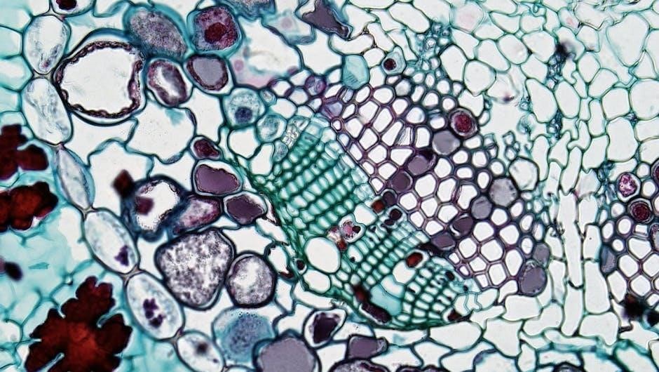
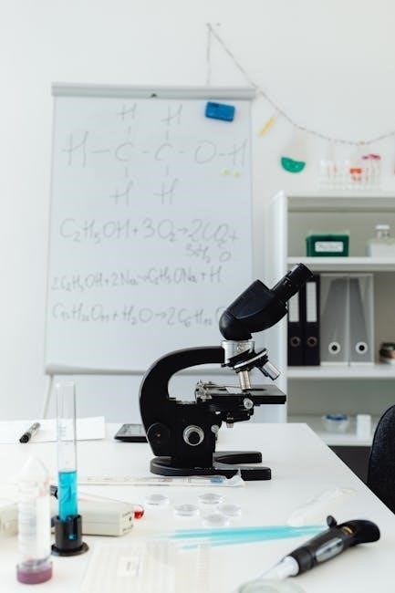
Methods for Studying Cellular Transport
Researchers employ diverse techniques to investigate the intricacies of cellular transport, gaining insights into mechanisms and identifying potential therapeutic targets. Tracer studies, utilizing labeled molecules, allow scientists to track the movement of substances across cell membranes and within cellular compartments, revealing transport rates and pathways.
Microscopy techniques, such as fluorescence microscopy and electron microscopy, provide visualization of transport proteins and vesicles. These methods enable observation of transport events in real-time and at high resolution.
Furthermore, sophisticated biochemical assays and genetic manipulation techniques are used to characterize transport protein function and identify genes involved in transport processes, furthering our understanding of cellular dynamics.
Tracer Studies
Tracer studies represent a cornerstone in unraveling the complexities of cellular transport, employing labeled molecules to meticulously track substance movement. These ‘tracers,’ often radioisotopes or fluorescent dyes, mimic the behavior of the molecules being studied, allowing researchers to chart their journey across cell membranes and through intracellular spaces.
By quantifying tracer uptake, distribution, and efflux, scientists can determine transport rates, identify specific transport pathways, and assess the influence of various factors like temperature or inhibitors. This technique provides invaluable data on the efficiency and selectivity of transport mechanisms.
The use of tracers allows for a dynamic view, revealing how cells adapt transport processes to changing conditions, ultimately enhancing our comprehension of cellular physiology.
Microscopy Techniques
Microscopy plays a pivotal role in visualizing and analyzing cellular transport processes, offering insights into the localization and dynamics of transport proteins and vesicles. Techniques like fluorescence microscopy, utilizing fluorescently labeled molecules, allow researchers to track the movement of specific substances within cells in real-time.
Confocal microscopy enhances resolution, enabling detailed observation of intracellular structures and transport pathways. Advanced techniques, such as super-resolution microscopy, push the boundaries of resolution even further, revealing previously unseen details of transport mechanisms.
Electron microscopy provides ultra-structural information, visualizing transport vesicles and membrane structures with exceptional clarity, contributing significantly to our understanding of cellular transport at the nanoscale.
Future Directions in Cellular Transport Research
Ongoing research focuses on developing more sophisticated tools to investigate the intricate mechanisms governing cellular transport, aiming to unravel the complexities of protein interactions and vesicle trafficking. A key area is the application of artificial intelligence (AI) to analyze large datasets generated from transport studies, predicting transport pathways and identifying potential drug targets.
Researchers are exploring novel methods for manipulating transport processes, utilizing optogenetics and chemogenetics to control protein activity with high precision. Furthermore, advancements in nanotechnology promise the creation of nanoscale sensors to monitor transport events in real-time.
Understanding the link between transport defects and disease remains a crucial focus, paving the way for innovative therapeutic strategies targeting specific transport impairments.